
Fall 2022 Newsletter
President's Message
 Greetings ASMH members,
I hope everyone had a great summer. It was wonderful to see all of the old and new members after two years of not being able to attend an in-person Mohs conference. To those who were not able to attend, we missed you and hope to see you at the next meeting.
I would like to say thank you to the Annual Program Committee for all their hard work in organizing another fantastic meeting for all levels of Mohs experience. Thank you to all of our incredible speakers and workshop leaders for all your time and for sharing your knowledge with the rest of us. I would also like to thank all the amazing vendors who showed us all the newest products and equipment.
Thank you to our outgoing board members: Past President Mo Gagnot, President Dan Gong, Director Linda Cesario, and ACMS Representative Dr. Dan Eisen, who have given so much of their time to help our society grow and become the best it can be. I would like to welcome and congratulate the new board members: Vice President Camille McKay, Director Stephanie Petrow, and ACMS Representative Dr. Randall Proctor. I look forward to working with each of you.
The Distinguished Service Award recipient was Norma Anderson, a member since 2004. She has been a Mohs Tech Trainer for the ASMH for many years. She has also been on the program committee since 2019 and chaired the first ever virtual meeting in 2021. Congratulations Norma! Thank you for everything you do for our society.
Congratulations to the 2022 ACMS Annual Meeting Scholarship winners Zipporah Cassidy and first-time meeting attendee Jean Marie Downes, and to 2020 ACMS Annual Meeting Scholarship winner, first-time meeting attendee Nicole Ryan. Scholarship recipients receive up to $1,000 to cover their annual meeting expenses, including registration, hotel, and travel. This scholarship is available to all members in good standing for at least 2 years. Each recipient must submit an article for the Summer newsletter highlighting their experience at the meeting.
The results are in for the 2021 Workforce Survey and there is a link to the article following. Please take the time to review the workforce survey.
View the 2021 Workforce Survey
The Mohs Tech Training Program will be having more training sessions in the new year. This is a great way to learn new skills and to learn the procedures and regulations. The ASMH is also looking for some new trainers. If interested, please contact Linda Cesario or Dan Gong.
There are many different ways to get more involved with our society. We are always looking for volunteers to write articles for our newsletter, present at the annual meeting, and/or sit either on the Board or on one of the many committees. If you are interested in volunteering, please email me or info@mohstech.org.
Sincerely,
Lindsey E. Riggs, HTL
President, 2022-24
Our New Executive Director
Lindsey E. Riggs, HTL
President, 2022-24
I am very happy to announce that Emily Knippel is our new Executive Director.
Emily has been with Executive Director, Inc. for more than eight years and she is very familiar with the ASMH having served in various roles with the American College of Mohs Surgery. She has a wealth of knowledge about association management and has extensive experience planning meetings and events and managing complex projects.
I believe that Emily is a great addition to the ASMH, and I am confident that she will help us accomplish great things. Please join me in welcoming her to this new role. She can be reached at eknippel@mohstech.org
ASMH Mohs Technician Training Program
Linda R. Cesario, DPM, HT (ASCP)
Director, Mohs Technician Training Program
The last ASMH Mohs Tech Training in June 2022 was held at Mercedes Scientific which was a sold-out event, bringing techs from all over the country and world to learn, sharpen their skills and network with each other. There were international attendees from as far away as Singapore! Miggy the Mohs Piggy, who adorns a lab coat with the ASMH logo, was also present and was a huge hit with the attendees as well as the faculty.
All attendees received 12 CEUs through the NSH and a certificate of attendance upon completion of the trainings bearing the distinguished logos of the ASMH and the ACMS. We are the only Mohs training program approved by the Mohs College.
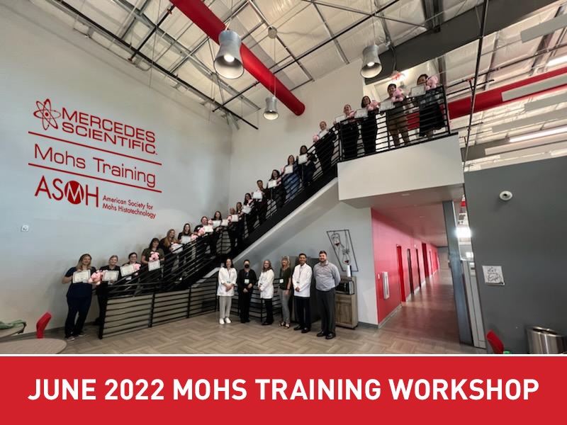
Thank you to Mercedes Scientific for graciously hosting the ASMH Mohs Tech Training Program and helping to promote education, a priority and mission of the ASMH.
Please check the ASMH website for more details on future trainings.
Toluidine Blue vs. H & E
By Reza Afzalneia, Memorial Sloan Kettering Cancer Center, Hauppauge, New York
Introduction
Tissue staining on frozen sections is one of the most important steps in obtaining an accurate pathologic diagnosis. Visualization and discernment of microscopic details can become difficult if staining is done improperly. Here, I will review H&E and Toluidine blue, the two most common stains used in the Mohs Labs, compare their advantages and limitations, and outline best lab practices in achieving optimal staining results.
H&E
H&E is the most widely used of all the various staining methods available in the frozen section. H&E is simple to perform, inexpensive and reliable. The two main dye components are hematoxylin and eosin. Hematoxylin is a natural dye derived from the Haematoxylon campechianum logwood tree, a tree native to Campeche’s Mexican state. It is a basic dye that stains acidic cell components such as nucleic acids, glycosaminoglycans, and acid glycoproteins, into a blue-purple hue. Eosin is an acidic dye and serves as an excellent counterstain to hematoxylin that targets the cytoplasm of cells, specifically mitochondria, secretory granules, and collagen. It gives differing shades of red and pink to the cytoplasm of different types of cells and different types of connective tissues. H&E can be performed in two different methods: progressive or regressive. In the progressive method, the tissue is initially stained with a hematoxylin solution that contains an excess of aluminum salts or acid, which increases nuclei affinity. Washing with water follows before a counterstain is applied. Variation in color is due to the selective affinity of tissue components to hematoxylin. With the regressive method, overstaining the tissue section with a neutral hematoxylin solution is the initial step. An acid alcohol is then used to remove excess stain, followed by an alkaline solution to achieve a neutralized tissue section. Stain intensity is controlled by visual examination with a microscope. The progressive method yields more reproducible results, but the regressive method offers a sharper degree of differentiation.
Advantages:
H&E yields excellent morphology, providing excellent contrast between cellular components. H&E provides better visualization of individual cell necrosis, keratinization, and keratin pearls, which are frequent histologic findings in well-differentiated squamous cell carcinoma. H&E offers stability of stained tissue lasting years without fading. H&E stain is also compatible when combined with various oxidants, mordants, and differentiating agents. H&E is reliable and simple to perform.
Limitations:
One of the main limitations of H&E is the length of the staining procedure. It can take from 3 to 5 minutes or longer to prepare one slide, depending on practice protocol. Due to the number of stains used, performing H&E during a frozen section can sometimes become challenging especially when multiple parts or sections are processed at once. H&E also poorly stains elastic material, reticular fibers, basement membranes, and lipids. It only provides limited insight into the specific biochemical nature of the tissue, specifically only the acidic and basic components.
Toluidine blue
Tolonium chloride, aka Toluidine blue (TB), is a cationic (basic) thiazine metachromatic dye which has a high affinity for acidic tissue components and turns nucleic acid blue and polysaccharides purple. When it binds to glycosaminoglycans, it gives off a reddish purple staining called metachromasia. This property is particularly useful in Mohs surgery for basal cell carcinoma (BCC) because the contrast of BCC’s blue aggregates with the reddish purple metachromasia of glycosaminoglycans in the surrounding stroma helps to delineate very small nests or a few clusters of tumor cells from adnexal structures.
Advantages:
Toluidine blue staining is faster and simpler than H&E and requires no counterstain. TB is a reliable stain and requires only one primary staining agent as opposed to H&E. Minimizing the staining time reduces patient waiting, facilitates patient flow, and frees up staff to perform other tasks. The metachromasia in BCCs and microcystic adnexal carcinoma (MAC) can be helpful in detecting small numbers of infiltrating tumor cells that otherwise might be missed. Mast cells and mucopolysaccharides in the stroma surrounding BCC may be highlighted in a magenta hue, prompting a diligent search for residual tumor. Mast cells have bright purple metachromatic granules when stain with TB and are abundant in the stroma of BCCs. Abundant mast cells may be indicative of residual tumor.
Limitations:
Nuclear detail is inferior compared to H&E. Many pathologists and Mohs surgeons are not trained with TB and therefore may find it difficult to use at first. It may take a while for them to become familiar with the staining pattern of TB. As with H&E, elastic material, reticular fibers, nerve fibers, and fat are difficult to identify.
Best Lab Practices
Whatever is the preferred staining method for the frozen section, the following general best lab practices help to ensure an optimal staining result.
- Wear appropriate personal protective equipment to avoid injury and cutaneous absorption.
- Optimal staining is achieved with 5-6 µm thick sections.
- Do not allow frozen sections to air-dry at any time during the staining procedure for better preservation of tissue morphology.
- Drain slides after each step to avoid injury and cutaneous absorption.
- Change staining solutions on a regular basis according to acceptable laboratory protocol. Consideration must be given to number of slides stained as well as time in use.
- Store chemicals in a cool, dry, well-ventilated area, and flammables in an approved cabinet or room.
- Ensure adequate ventilation and regularly check fume contaminant hoods that can minimize the amount of inhaled chemical vapor.
- Quality control should be performed daily before proceeding with patient specimens.
Common problems with rapid H&E staining
Weak Hematoxylin staining:
Due to autolysis or poor fixation, over-decalcification, inadequate staining time, excessive de-staining, weak hematoxylin due to carryover, contaminants, thin sections, and inadequate removal of alcohol or insufficient pre-rinsing with water prior to staining with hematoxylin.
Excessive Hematoxylin staining:
Due to drying of tissue, excessive staining times, too weak or inadequate de-staining time, thick section.
Weak Eosin staining:
Due to too high Eosin pH, contaminant in the alcohol rinse, deteriorating eosin due to excessive carryover, thin sections, inadequate staining time, over-differentiation by subsequent 95% alcohol.
Excessive Eosin staining:
Due to stronger dye solution (can be due to excessive evaporation), use of isopropyl alcohol as the rinsing agent, thick sections, excessive staining times Water beads or white haze under the coverslip: Due to incomplete dehydration of the section, formed by mixture of water and the clearing agent.es
Eosin bleeding from the tissue section:
Due to inadequate clearing of alcohol, which will remain within the tissue section and cause the bleeding.
Common problems with Toluidine blue
Weak Toluidine blue staining:
Due to autolysis or poor fixation, over-decalcification, inadequate staining time, excessive de-staining/rinsing with water, weak solution due to water carryover, contaminants, thin sections
Excessive Toluidine blue staining:
Due to drying of tissue, excessive staining times, inadequate de-staining/rinsing with water, thick sections
Water beads or white haze under the coverslip:
Due to incomplete dehydration of the section, formed by mixture of water and the clearing agent.
Sample Toluidine blue staining method:
Alcoholic formalin (10 dips)
Tap water (10 dips)
Tap water (10 dips)
1% T-blue (40 seconds)
Tap water (10 dips)
70% ethanol (10 dips)
95% ethanol (10 dips)
100% ethanol (10 dips)
100% ethanol (10 dips)
Clearing agent (10 dips)
Clearing agent (10 dips)
Coverslip with compatible mounting medium
Conclusion
Preference for the staining method in the frozen section is usually based on individual training or personal experience. The advantages and limitations of each technique should be considered, whether the preference is H&E or TB. It is important to recognize that staining characteristics vary with technique, brand of the solution/stain, and experience. Firm understanding of the principles of the H&E or TB staining methods is crucial in troubleshooting for problems. Tissue staining should be reproducible and reliable. Excellent visualization and discrimination of histologic detail of tissue sections are crucial in a rapid and accurate pathologic diagnosis in frozen section.
References
- Aslam A, Aasi SZ. Frozen-Section Tissue Processing in Mohs Surgery. Dermatol Surg. 2019 Dec;45 Suppl 2:S57-S69. doi: 10.1097/DSS.0000000000002260. PubMed PMID: 31764292.
- Bancroft JD, Gamble M, editors. Theory and Practice of Histological Techniques. 6th ed. New York: Churchill Livingstone;2008.
- Bancroft JD, Stevens A. Histopathological Stains and Their Diagnostic Uses. New York: Churchill Livingstone; 19
- Carson, Freida L., and Christa Hladik Cappellano. Histotechnology: A Self-instructional Text. 4th ed. Chicago: ASCP Press, 2015. 120-121
- Humphreys TR, Nemeth A, McCrevey S, Baer SC, Goldberg LH. A pilot study comparing toluidine blue and hematoxylin and eosin staining of basal cell and squamous cell carcinoma during Mohs surgery. Dermatol Surg. 1996 Aug;22(8):693-7. PubMed PMID: 8780761.
- Junqueira LC, Carneiro J. Basic Histology. 11th ed. New York: McGraw Hill, Inc.; 2005.
- Ross MH, Wojciech P. Histology: A Text and Atlas. 5th ed. Baltimore: Lippincott Williams & Wilkins; 2006.
- Larson K, Ho HH, Anumolu PL, Chen TM. Hematoxylin and eosin tissue stain in Mohs micrographic surgery: a review. Dermatol Surg. 2011 Aug;37(8):1089-99. doi: 10.1111/j.1524-4725.2011.02051.x. Epub 2011 Jun 2. Review. PubMed PMID: 21635628.
- Luna L, editor. Manual of Histologic Staining Methods of the Armed Forces Institute of Pathology. 3rd ed. New York: McGraw-Hill, Inc.; 1968.
- Preece A. A Manual for Histologic Techniques. 3rd ed. Boston: Little, Brown, and Company; 1972.
- Sridharan, G; Shankar, AA (2012). Toluidine Blue: A review of its chemistry and clinical utility. J Oral Maxillofac Pathol. 16: 251–5. doi:10.4103/0973-029X.99081.
- Tehrani H, May K, Morris A, Motley R. Does the dual use of toluidine blue and hematoxylin and eosin staining improve basal cell carcinoma detection by Mohs surgery trainees? Dermatol Surg. 2013 Jul;39(7):995-1000. doi: 10.1111/dsu.12180. Epub 2013 Mar 6. PubMed PMID: 23465179.
- Todd MM, Lee JW, Marks VJ. Rapid toluidine blue stain for Mohs’ micrographic surgery. Dermatol Surg. 2005 Feb;31(2):244-5. PubMed PMID: 15762224.
- Wang SQ, Goldberg LH, Nemeth A. The merits of adding toluidine blue-stained slides in Mohs surgery in the treatment of a microcystic adnexal carcinoma. J Am Acad Dermatol. 2007 Jun;56(6):1067-9. PubMed PMID: 17504725.
- Wittekind D. Traditional staining for routine diagnostic pathology including the role of tannic acid. 1. Value and limitations of the hematoxylin-eosin stain. Biotech Histochem 2003;78:261-70.
- Carson, Freida L., and Christa Hladik Cappellano. Histotechnology: A Self-instructional Text. 4th ed. Chicago: ASCP Press, 2015. 120-121
Annual Meeting Scholarship Article
By Jean Marie Downes, HTL, Innovative Dermatology, Chicago, IL
Every year since I began in the field of Mohs, I had a full weekend off in May due to the annual Mohs meeting taking place across the country. After 12 years, I was finally able to attend the meeting this year in Philadelphia and I could not be more pleased.
I have been to various conferences or educational meetings in the past, but this was truly the most informational and time well spent. The meeting was run by board members and almost all the presentations were given by fellow histotechs, and this is where the information really settled in. The board did a wonderful job of lining up several topics that each of us encounter every day. Not only did I learn new techniques, tricks, and Mohs facts, but I also met numerous techs who made me feel welcome.
My favorite portion of the meeting was the hands-on advanced cryostat training. It gave me an opportunity to ask questions and see other techs perform, and those skills have since been put into practice. In addition to providing intellectual information, the accommodations, events planned, and meals provided were top notch. I left Philadelphia inspired and humbled to know that I am a part of such a wonderful community and that we all share the same love of Mohs. I encourage all technicians to attend the meeting every year – it will change the way you look at Mohs.
Highlights of the 2022 Annual Meeting
By Melinda Chow, MS, HT (ASCP) CM, Memorial Sloan Kettering Cancer Center, Basking Ridge, NJ
After two years of not being able to meet in person due to the Covid-19 stricken years of 2020 and 2021, the 2022 Annual Meeting took place at the Marriott Hotel in downtown Philadelphia, a.k.a, the City of Brotherly Love. Over 160 vaccinated attendees gathered for the 3 days of informative pre-conference workshops, myriad talks on interesting topics, networking, and an opportunity to explore the attractions the city has to offer and visit the many historic landmarks.
The pre-conference workshops covered topics ranging from beginner to advanced cryostat use. Attendees learned the technique of cutting frozen sections from experienced Mohs leaders and learned how to stain frozen sections for immunohistochemistry. One could also learn the theory and procedures for routine H & E staining of Mohs Slides; how to prepare and pass CAP, CLIA inspections to regulatory standards and the survey process. There were also workshops on how to relax and embed tissues using the slide method, and a workshop on technical guidance on potential pitfalls in the Mohs procedures. This workshop encompassed the common artifacts seen in the Mohs laboratories and how to best troubleshoot and avoid them.
This year’s program consisted of a wide range of topics. Immunohistochemistry, Mart-1 staining and the new melanoma antibody marker, PRAME have sparked interest as many questions were asked at the end of each talk. Camille McKay and her collaborators gave one workshop and three talks in this year’s meeting. Her workshop and talks mainly focused on the regulatory standards and the gray areas of the survey process with JACHO and CLIA. She also shared her thoughts on developing strong skillsets among Mohs techs and promoting the field of Mohs histotechnology. We look forward to her talks in the future.
A seasoned Mohs professional presented a novel technique of embedding specimens using rubber cement. This innovative technique could help eliminate some of the problems with embedding. It was an eye-opening opportunity to learn this new technique of embedding Mohs specimens.
Dr. K Connolly discussed a type of nonmelanoma skin cancer, dermatofibroscaroma protuberans. It is a type of soft tissue sarcoma found on the trunk, arms and legs in relatively young individuals between the ages of 20-50. Her talk covered the management of these larger specimens using new treatment and Mohs surgery. Dr. M Criscito talked about the challenges in the management of cutaneous squamous cell carcinoma with perineural invasion.
There was an instructional talk on how to write and submit a scientific paper. There was a video on MohsHacks that can help with your daily Mohs lab operation. A great presentation on the tips for doing whole mount sectioning using the reverse slide embedding method was also given on the second day of the scientific program.
The Q & A session in the Troubleshooting open forum was full of good questions. Questions on embedding and staining dominated this session. This session was very well attended.
A tech from Tech One Biomedical gave an entertaining and informative talk on cryostat quick fixes. Some useful tips on troubleshooting cryostat problems were presented in this talk. His YouTube videos can also be found on the internet.
There were other topics on margin assessment in Mohs surgery, working with difficult tissues, embedding pitfalls in Mohs processing and communication in the Mohs team. All these topics are of paramount importance, informative, educational and inspiring to the Mohs techs. Whether you are a novice in the field or a seasoned Mohs professional, you surely gained knowledge by attending this year’s annual meeting.
On a business note, Norma Anderson was the recipient of the 2022 Distinguished Service Award. Three scholarships were also awarded for the best abstract submissions.
This year’s annual meeting has been an overwhelming success. Everything went smoothly as planned and no positive Covid case was reported.
2023 ASMH Board of Directors Nominations Now Open
Nominations are now being accepted for two (2) positions on the Board of Directors. In early 2023, the membership will elect one (1) Secretary-Treasurer and one (1) Director for the 2023 term. Board terms will begin at the conclusion of the 2023 Annual Meeting. The Secretary-Treasurer will serve a two-year term concluding at the 2025 Annual Meeting. The Director will serve a three-year term concluding at the 2026 Annual Meeting. A description of the duties for these positions can be found here.
Nominations will be accepted until January 11, 2023 and should be sent, faxed, or e-mailed to the ASMH office. Self-nominations are accepted. Elections will take place via electronic voting which will begin in February 2023. To send a nomination or ask a question about the nomination process, contact info@mohstech.org.
Think about someone who you feel is qualified for these positions and ask them to serve – or nominate yourself. A nomination form, a well as a description of the duties for these two positions, can be found here.
Member Resources
2021 Workforce Survey
One of the benefits of ASMH membership is the ability to contribute to ASMH’s biennial Workforce Survey. Data obtained from this survey will not only help examine industry standards and trends, but will assist you and other Mohs technicians in determining if you are paid competitively. When you know what the industry standard is, you can negotiate salary with your current employer during the performance appraisal process. This information can also assist you with future job search efforts.
The 2021 Workforce Survey is now available on the Current Members page of the ASMH website.
ASMH Bibliography
The ASMH bibliography is your resource and we strongly encourage you to explore it. Efforts will be made to keep the articles up to date and relevant to current best practices while keeping the scope wide enough to interest the broadest of minds. It is our intention to cover subjects ranging from basic lab techniques, Mohs embedding, cryotomy, routine and immunohistochemical staining, lab safety, ethics and work flow. This is a continually evolving project that will be guided by your feedback, suggestions and more importantly, your contributions, so please share interesting articles and send topic suggestions to info@mohstech.org.
Go to the bibliography and find new COVID-19 resources: Laboratory Biosafety & Coronavirus Disinfection in Histopathology.
Member Marker

The American Society for Mohs Histotechnology has a rich tradition of dedicated members that work hard to keep the ASMH true to the original commission; to uphold the importance of training, collaboration and consistency, while working in conjunction with the American College of Mohs Surgery to ensure that the high standards set by Dr. Mohs continue to be met.
Members are encouraged to download, and use this graphic in your email signature, on your social media profile or added to your professional online presence. Please remember that you may not, in any way, alter, manipulate or change this graphic. Contact info@mohstech.org should you have any questions about proper use.
Note to ASMH Newsletter Readers:
Reference to any specific commercial product, process, or service by trade name, trademark, manufacturer, or otherwise throughout this e-newsletter, does not constitute or imply its endorsement, recommendation, or favoring by the American Society for Mohs Histotechnology. The views and opinions of authors expressed do not state or reflect those of the American Society for Mohs Histotechnology.